Category Medical Inventions and Discoveries

What is a speech generating device?

An electronic device, it is of great use to those with difficulty in speaking. How does it work?

A speech generating device (SGD) is an electronic device that creates speech for those who have difficulty in speaking. Most SGDS are connected to a keyboard, eye sensor or other such keyboard input device that allows the user to select the words to be spoken. The user can enter words or phrases with or use a visual display with images to produce speech.

Digitally recorded human voices speaking actual words are stored in the device and played back upon selection. A variety of voices to match a users gender and age are available. Some SGDS also use computer generated speech similar to the ones used in automated telephone systems.

SGDS have certain advantages over sign boards or other communication methods. It enables a person with speech impairment to communicate through spoken words.

This means the user can easily draw the attention of someone at a distance or sitting in another room or even talk on the phone! SGDS are very effective for autistic children with limited speech ability. World renowned scientist Stephen Hawking used speech generating devices for years. He used to prepare his lectures at the Cambridge University in advance and deliver them using the SGD.

Picture Credit : Google

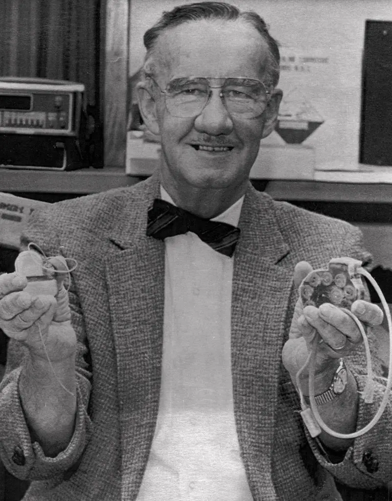
How did Wilson Greatbatch invented the pacemaker?

Quite by accident, American engineer Wilson Greatbatch invented the implantable cardiac pacemaker in the year 1958. Read on to know how….

An artificial pacemaker is a small battery-operated electronic device that’s placed under the skin in the chest to help control the heartbeat. The first successful implantable pacemaker was invented in 1958 by an American electrical engineer, Wilson Greatbatch. He was making a heart rhythm recorder when, by mistake, he added a wrong electronic component. He was shocked when the device, instead of simply recording the sound of the heartbeat, produced electronic pulses quite similar to the sounds made by a healthy heart.

It struck him then that the device could make an unhealthy heart beat in rhythm by delivering electrical pulses to make the heart muscles contract and pump blood. For two years he worked on modifying the device. He miniaturised it, coated it with a kind of resin to prevent it from getting damaged by body fluids, and powered it with a mercury-zinc battery.

Greatbatch discussed his invention with surgeon William Chardack, whom he met in a chance encounter. In 1960, the Chardack-Greatbatch pacemaker was implanted in an elderly man with an irregular heartbeat. The patient’s life was extended by 18 months.

Pacemakers today are about the size of a bullet. They are encased in titanium and keep the heart ticking with regular beats through computer-guided electrical pulses. They are inserted through the leg up into the right ventricle. Their batteries can last from 5 to 15 years.

Picture Credit : Google 

WHAT IS CRYOGENIC ELECTRON MICROSCOPY?

Did you know scientists hope to develop targeted drugs for hard-to-treat diseace using the technique?

Ultra high-definition 3D videos of cells inside the body taken by an advanced microscope are creating a revolution in modern biology. Previously, transmission electron microscope (TEM) images of living cells (called biomolecules) were indistinct blobs. This was because the high-energy electron beams would dry out the water surrounding the molecules and burn them.

In cryogenic electron microscopy (cryo-EM), biomolecules are cooled to extremely low temperatures and embedded in vitrified water (ice that has no crystals). This ensures that they are preserved intact. The three scientists who developed the cryo-EM technique won the 2017 Nobel Prize in Chemistry.

The current level of sophistication in the cryo-EM technique is largely due to advances in camera technology, image processing and computer software. The biomolecules are photographed from thousands of different angles and at different stages. The images are put together to create a video, allowing researchers to see the structure of the molecules in live action within the cell.

Using this technique, scientists hope to develop targeted drugs for hard-to-treat diseases such as dementia and Parkinson’s.

Picture Credit : Google