Category Human Body

What is ligament?

Ligaments are made out of connective tissue that has a lot of strong collagen fibers in it. They are found in different shapes and sizes in the body. Some look like pieces of string, others look like narrow or wide bands. There are arch-shaped ligaments, too.

Ligaments often connect two bones together, particularly in the joints: Like strong, firmly attached straps or ropes, they stabilize the joint or hold the ends of two bones together. This ensures that the bones in the joint don’t twist too much or move too far apart and become dislocated.

But there are also some ligaments that aren’t connected to bones. For instance, some make sure that internal organs are kept in place. A typical example is the womb, which is kept in the right position in the pelvis by ligaments. Ligaments may also connect two or more organs to each other. For instance, the liver, intestine and stomach are held in place by ligaments in the abdominal cavity. These ligaments often have sensitive structures like blood vessels or gland ducts running through them. The strong connective tissue in the ligaments protects these structures and prevents them from bending, twisting or tearing.


Picture Credit : Google

Where are Ossicles found?

The ossicles are situated in the middle ear and suspended by ligaments. They articulate with each other through synovial joints to form a chain across the length of the middle ear from the tympanic membrane (laterally) to the oval window (medially). The ossicles transmit mechanical vibrations of the tympanic membrane across this chain to the oval window where fluids of the inner ear will move and excite receptors. This process allows sound to be transformed into electrical signals which are then sent to the brain.  This article will explore the function of the auditory ossicles, their bony features, articulations, associated muscles, and some clinical aspects.

Their role is to mechanically amplify the vibrations of the tympanic membrane and transmit them to the cochlea where they can be interpreted as sound. They are located in the middle ear cavity and articulate with each other via two tiny synovial joints. The stapes also articulates with the oval window via the stapediovestibular joint, which is a syndesmosis 3; this joint transmits the ossicular vibrations to the endolymph in the vestibule.

Interestingly, they are the only bones in the body that do not grow after birth, and are also the smallest bones in the body (variant tiny sesamoids aside).


Picture Credit : Google

What is bone marrow?

Bone marrow is the spongy tissue inside some of the bones in the body, including the hip and thigh bones. Bone marrow contains immature cells, called stem cells.

Most red blood cells, platelets, and most of the white blood cells are formed in the red marrow. Yellow bone marrow produces fat, cartilage, and bone.

White blood cells survive from a few hours to a few days, platelets for about 10 days, and red blood cells for about 120 days. These cells must be constantly replaced by the bone marrow, as each blood cell has a set life expectancy.

Certain conditions may trigger additional production of blood cells. This may happen when the oxygen content of body tissues is low, if there is loss of blood or anemia, or if the number of red blood cells decreases. If these happen, the kidneys produce and release erythropoietin, a hormone that stimulates the bone marrow to produce more red blood cells.

The bone marrow also produces and releases more white blood cells in response to infections, and more platelets in response to bleeding. If a person experiences serious blood loss, yellow bone marrow can be activated and transformed into red bone marrow.

Healthy bone marrow is important for a range of systems and activities.


Picture Credit : Google

Which is the longest and strongest bone in the human body?

The femur bone is the strongest and longest bone in the body, occupying the space of the lower limb, between the hip and knee joints.

The femur is an integral component of ambulation. A lot of the large thigh muscles arise from and insert on the various parts of the femur. Muscles that originate from the pelvis and insert on the anterior or posterior surface of the femur to facilitate flexion and extension around the hips. Muscles which arise from the femur will cross the knee joint to insert on the proximal tibia promote flexion and extension around the knee. The tables below summarize the thigh muscles and their points of origin or insertion with respect to the femur.

Important features of this bone include the head, medial and lateral condyles, patellar surface, medial and lateral epicondyles, and greater and lesser trochanters. The head is where the bone forms the hip joint with the innominate bone. The condyles are the points of articulation (connection) with the tibia, which is a lower leg bone. The patellar surface is the groove where the bone adjoins with the patella, or kneecap. The epicondyles and trochanters are all important attachment sites for various muscles.


Picture Credit : Google

What is Hyoid?

The hyoid bone is a ‘U’ shaped structure located in the anterior neck. It lies at the base of the mandible (approximately C3), where it acts as a site of attachment for the anterior neck muscles.

The hyoid consists of a body, two greater horns, and two lesser horns. The body forms the central quadrilateral-shaped broad segment of the hyoid. The greater horns are larger and longer than the lesser horns of the hyoid. The greater horns and lesser horns are also known as cornu majus and cornu minus, respectively. The body and the greater horns appear to give the hyoid its U-shape with the greater horns forming the limbs of the “U” on either side of the body. The greater and lesser horns normally unite to the body of the hyoid via fibrous tissue or a true joint. As age progresses, there is a physiological progression of ankylosis of the joints connecting the greater and lesser horns with the body of the hyoid.  

The hyoid takes part in all possible functional actions of the orofacial complex. It preserves the patency of the airway between the oropharynx above and tracheal rings below. It also connects to the larynx and hence plays a role in phonation. Other functions include tongue movement, mastication, swallowing, prevention of regurgitation, and even respiration. Furthermore, the hyoid maintains the posture of the head, due to the complex connection it presumes between the mandible and the cervical spine.


Picture Credit : Google

Why is food nutrition important?

Nutrition is the food your body needs to grow, move, and keep all its parts working. The body can make some of the substances it needs, but the rest have to come from the food we eat. The digestive system breaks food down into simple chemicals called nutrients that the body can use. Nutrients energize the cells ready for work, provide material for new tissues, and help to repair injuries.  

Good nutrition is an important part of leading a healthy lifestyle. Combined with physical activity, your diet can help you to reach and maintain a healthy weight, reduce your risk of chronic diseases (like heart disease and cancer), and promote your overall health.

The link between good nutrition and healthy weight, reduced chronic disease risk, and overall health is too important to ignore. By taking steps to eat healthy, you’ll be on your way to getting the nutrients your body needs to stay healthy, active, and strong. As with physical activity, making small changes in your diet can go a long way, and it’s easier than you think!


Picture Credit : Google

What is inside the stomach?

The stomach’s wall has three muscle layers that run in different directions. During digestion, these contract in turn to churn food while mixing it with acidic gastric juice. Thick mucus stops gastric juice from damaging the stomach’s own delicate lining.
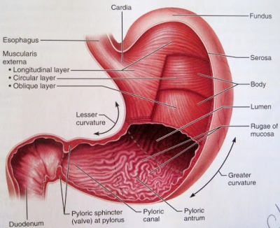
Pylric sphincter

Normally closed to keep food in the stomach, this ring of muscle opens slightly once food has been processed to allow a controlled flow or chyme into the duodenum.

Duodenum

The duodenum is the first of the three parts of the small intestine that receives partially digested food from the stomach and begins with the absorption of nutrients. It is directly attached to the pylorus of the stomach. The first part of the small intestine is about 25 cm (10 in) long.

Gastric mucosa

The stomach’s inner layer contains gastric glands. The mucosa is always covered by a layer of thick mucus that is secreted by tall columnar epithelial cells. Gastric mucus is a glycoprotein that serves two purposes: the lubrication of food masses in order to facilitate movement within the stomach and the formation of a protective layer over the lining epithelium of the stomach cavity. 

Oesophagus

Food is carried in this tube from the throat to the stomach. The upper part of the oesophagus is behind the windpipe (trachea). The windpipe is the tube that connects your mouth and nose to your lungs, so you can breathe. Below your lungs is a layer of muscle called the diaphragm. It helps you to breathe. Most of your oesophagus sits above the diaphragm in your chest.

Serous layer

The stomach is covered by this protective layer. Serous membranes have two layers. The parietal layers of the membranes line the walls of the body cavity (pariet- refers to a cavity wall). The visceral layer of the membrane covers the organs (the viscera). Between the parietal and visceral layers is a very thin, fluid-filled serous space, or cavity.

Longitudinal muscle

This layer runs the length of the stomach. This layer is composed of smooth muscle, continuous with the smooth muscle which surrounds the esophagus. Below this longitudinal muscle is the Auerbach’s plexus, or myenteric plexus, above the middle circular. 

Circular muscle

This layer wraps around the stomach. It wraps in a circular orientation around the pylorus, and is held in a constricted state normally. The normal constriction of this muscle is what creates the pyloric sphincter, which controls the movement of chyme into the duodenum. This layer is concentric to the longitudinal axis of the stomach. 

Oblique muscle

This layer runs diagonally.  It wraps around the body of the stomach, extending downward to form the pyloric sphincter along with the circular muscle layer. This layer is responsible for creating the motion that churns and physically breaks down the food. It is the only layer of the three which is not seen in other parts of the digestive system. 

Gastric pits

These pits lead to the gastric glands, which make and release gastric juice. This liquid contains a mixture of enzymes, hydrochloric acid, and mucus.

Rugae

Deep folds in the stomach wall disappear when it stretches, as it fills the food. The inner layer of the stomach is full of wrinkles known as rugae (or gastric folds). Rugae both allow the stomach to stretch in order to accommodate large meals and help to grip and move food during digestion.

Protective coat

Thick fluid coats and lining, preventing the stomach from being digested by its own gastric juice. The mucus protects the gastric mucosa from auto digestion by e.g. pepsin and from erosion by acids and other caustic materials that are ingested. Bicarbonate ions, secreted by the surface epithelial cells.


Picture Credit : Google

What is the function of lungs?

The two lungs take up most of the space in the chest. Their key function is to get oxygen into, and waste gases out of, the bloodstream. That oxygen is used by the body’s cells to release energy, a process that produces waste carbon dioxide.

Breathing draws air rich in oxygen into the lungs through the airways, then pushes air containing carbon dioxide in the opposite direction. Lungs are spongy because they are packed with branching, air-filled tubes that get narrower and narrower before ending in tiny air sacs (alveoli). It is here that oxygen is swapped for carbon dioxide.

The lungs are like bellows. As they expand, air is sucked in for oxygen. As they compress, the exchanged carbon dioxide waste is pushed back out during exhalation.

When air enters the nose or mouth, it travels down the trachea, also called the windpipe. After this, it reaches a section called the carina. At the carina, the windpipe splits into two, creating two mainstem bronchi. One leads to the left lung and the other to the right lung.

From there, like branches on a tree, the pipe-like bronchi split again into smaller bronchi and then even smaller bronchioles. This ever-decreasing pipework eventually terminates in the alveoli, which are little air sac endings.


Picture Credit : Google


How blood clots?

After a cut, blood seeps from the wound, triggering an immediate repair process. The blood cells take action immediately. They stop the leak, form a plug, and destroy harmful bacteria. A scab forms, and the clot dissolves when the wound has healed.

Injury

A cut in the skin damages blood vessels. Platelets start to group together at the site of the injury.

Plug

The platelets release chemicals that make fibrin, a sticky thread-like protein. Red cells get stuck in the threads, forming a plug. White blood cells arrive to hunt for germs.

Clot

The fibrin threads contract, binding red blood cells and platelets together in a sticky clot, which closes the wound.

Scab

The clot near the skin’s surface dries out to form a protective scab, which covers the healing wound.


Picture Credit : Google

What is the function of ribs and chest muscles?

The chest, or thorax, lies between the neck and the abdomen. Inside the thorax lie the heart, lungs, and major blood vessels. The ribcage surrounding them is formed by the backbone, ribs, costal cartilages, and sternum (breastbone).

The ribcage is strong enough to protect the vital organs, but flexible enough to expand and contract for breathing. Attached to the ribcage are the muscles of the chest. Together with the diaphragm, many of these muscles help with breathing.

They move superiorly, inferiorly, anteriorly and posteriorly to facilitate breathing (their flexibility in their movement increases/decreases the size of the thoracic cavity; assisting the lungs in respiration. Control of these movements is via the diaphragm, external intercostals, and the intercartilaginous portion of the internal intercostals). They play a role in erythropoiesis during development (at birth, the erythropoiesis sites change, it recedes in long bones and persists in flat bones, like ribs).


Picture Credit : Google