Category Human Body

Which part of the brain connects the cerebrum and cerebellum to the spinal cord?

Brainstem: acts as a relay center connecting the cerebrum and cerebellum to the spinal cord. It performs many automatic functions such as breathing, heart rate, body temperature, wake and sleep cycles, digestion, sneezing, coughing, vomiting, and swallowing.

The brain stem has 3 areas:

  • midbrain (also called the mesencephalon)
  • pons
  • medulla oblongata

The brain stem sends information to and from the other parts of the brain to the rest of the body and controls:

  • breathing
  • body temperature
  • blood pressure
  • heart rate
  • hunger and thirst
  • digestion of food

More specific functions of each part of the brainstem are dependent of the cranial nerve nuclei and tracts that reside in each area. The medulla oblongata controls the respiratory function, cardiovascular system, as well as gastrointestinal and digestive activities. The pons is involved in controlling movements of the body and equilibrium. Finally, the midbrain controls eye movements and integrates it with auditory input. 


Picture Credit : Google

What happens after a stroke in the cerebellum?

Before we discuss the various cerebellar stroke effects, it’s important to understand the function and structure of the cerebellum itself. The cerebellum — which in Latin means “little brain” — is the second largest part of the brain, following the cerebrum. The cerebellum is part of the hindbrain, located along the rear base of the skull and tucked just beneath the cerebrum. Although the cerebellum only accounts for roughly 10 percent of the brain’s volume, it contains more than half of its total neurons. This mighty “little brain” is responsible for controlling and regulating balance and coordination. The cerebellum plays a major role in fine motor movements and especially muscle memory. In other words, it controls the movements we learn over time through repetition, such as playing an instrument or throwing a baseball.

After a stroke occurs, immediate medical attention is vital to minimizing neural damage. Signs to look for to determine if a patient has suffered a cerebellar stroke include headache, nausea, loss of coordination, vomiting, and difficulty swallowing. Dizziness is one of the most commonly reported cerebellar stroke symptoms, with nearly three-quarters of stroke patients experiencing instances of vertigo, the sensation of falling to the left or right. Other cerebellar stroke effects such as double-vision or nystagmus (condition involving involuntary eye movements) can exacerbate instances of vertigo. Nausea, with or without vomiting, is also extremely common, affecting more than half of cerebellar stroke patients.


Picture Credit : Google

What is a haemorrhage?


Bleeding, also called hemorrhage, is the name used to describe blood loss. It can refer to blood loss inside the body, called internal bleeding, or to blood loss outside of the body, called external bleeding.

Blood loss can occur in almost any area of the body. Internal bleeding occurs when blood leaks out through a damaged blood vessel or organ. External bleeding happens when blood exits through a break in the skin

When blood from trauma irritates brain tissues, it causes swelling. This is known as cerebral edema. The pooled blood collects into a mass called a hematoma. These conditions increase pressure on nearby brain tissue, and that reduces vital blood flow and kills brain cells.

Bleeding can occur inside the brain, between the brain and the membranes that cover it, between the layers of the brain’s covering or between the skull and the covering of the brain.


Picture Credit : Google

Which is the risk factor of stroke?

Lifestyle factors that increase your risk of stroke include high blood pressure, smoking, diabetes, high blood cholesterol levels, heavy drinking, high salt and high fat diet and lack of exercise. Someone who has already experienced a stroke is at increased risk of having another.

High blood pressure (hypertension) is the most significant risk factor for stroke. Blood pressure refers to the pressure inside the arteries. Normal blood pressure is around 120/80, while high-normal blood pressure is 120/80 to 140/90. High blood pressure is when your blood pressure is consistently over 140/90. This is called ‘hypertension’.

Hypertension means that the blood is exerting more pressure than is normal or healthy. Over time, this weakens and damages blood vessel walls, which can lead to stroke, particularly cerebral haemorrhage.

Hypertension may also cause thickening of the artery walls, resulting in narrowing and eventual blockage of the vessel (ischaemic stroke). In atherosclerosis, the pressure of your pumping blood could ‘hose off’ debris from damaged artery walls. The circulating debris (called emboli) can cause a stroke by lodging in and blocking a blood vessel in the brain.


Picture Credit : Google

Why does brain stroke happen?

A stroke, or “brain attack,” occurs when blood circulation to the brain fails. Brain cells can die from decreased blood flow and the resulting lack of oxygen. There are two broad categories of stroke: those caused by a blockage of blood flow and those caused by bleeding into the brain. A blockage of a blood vessel in the brain or neck, called an ischemic stroke, is the most frequent cause of stroke and is responsible for about 80 percent of strokes. These blockages stem from three conditions: the formation of a clot within a blood vessel of the brain or neck, called thrombosis; the movement of a clot from another part of the body such as the heart to the brain, called embolism; or a severe narrowing of an artery in or leading to the brain, called stenosis. Bleeding into the brain or the spaces surrounding the brain causes the second type of stroke, called hemorrhagic stroke.

The most common symptoms of a stroke are:

  • Weakness or numbness of the face, arm, or leg on one side of the body
  • Loss of vision or dimming (like a curtain falling) in one or both eyes
  • Loss of speech, difficulty talking, or understanding what others are saying
  • Sudden, severe headache with no known cause
  • Loss of balance or unstable walking, usually combined with another symptom


Picture Credit : Google

What is the importance of water in our body?

The body needs water to stay alive. Every cell, tissue, and organ relies on a regular water supply to function properly. Water makes up more than half of the body. It is found inside cells, as well as in blood and other fluids, such as lymph, tears, saliva, sweat, and urine. The brain constantly monitors water level inside the body so it can make sure it maintains the correct balance.

Adequate water intake enables your body to excrete waste through perspiration, urination, and defecation. Water helps your kidneys remove waste from your blood and keep the blood vessels that run to your kidneys open and filter them out, according to the National Kidney Foundation. Water is also important for helping prevent constipation, points out the University of Rochester Medical Center. However, as research notes, there is no evidence to prove that increasing your fluid intake will cure constipation.

Your body loses fluids when you engage in vigorous exercise, sweat in high heat, or come down with a fever or contract an illness that causes vomiting or diarrhea, according to the Centers for Disease Control and Prevention. If you’re losing fluids for any of these reasons, it’s important to increase your fluid intake so that you can restore your body’s natural hydration level. Your doctor may also recommend that you drink more fluids to help treat other health conditions, like bladder infections and urinary tract stones. If you’re pregnant or nursing, you may want to consult with your physician about your fluid intake because your body will be using more fluids than usual, especially if you’re breastfeeding.


Picture Credit : Google

What are glomeruli?

Inside the kidneys are thousands of tiny structures called glomeruli (one is called a glomerulus), working hard to filter toxic waste from the bloodstream. These tightly coiled clusters are the body’s smallest blood vessels (capillaries).

Each glomerulus works like a miniature sieve, transferring waste and excess water from the blood through the capillary wall into a renal tubule. This fluid drains away towards the bladder as urine.

The glomerulus receives its blood supply from an afferent arteriole of the renal arterial circulation. Unlike most capillary beds, the glomerular capillaries exit into efferent arterioles rather than venules. The resistance of the efferent arterioles causes sufficient hydrostatic pressure within the glomerulus to provide the force for ultrafiltration.

The glomerulus and its surrounding Bowman’s capsule constitute a renal corpuscle, the basic filtration unit of the kidney.[2] The rate at which blood is filtered through all of the glomeruli, and thus the measure of the overall kidney function, is the glomerular filtration rate.


Picture Credit : Google

What is dialysis?

If the kidneys become damaged or diseased, a dialysis machine can be used to filter the blood instead. This large machine takes up to four times longer than kidneys to clean the body’s blood – which shows what an efficient unit the kidneys are.

The dialysis machine acts as an artificial set of kidneys. Blood flows from the body to the machine, toxic waste and excess fluid are removed, and cleaned blood is returned.

Some kinds of acute kidney failure get better after treatment. In some cases of acute kidney failure, dialysis may only be needed for a short time until the kidneys get better.

In chronic or end stage kidney failure, your kidneys do not get better and you will need dialysis for the rest of your life. If your doctor says you are a candidate, you may choose to be placed on a waiting list for a new kidney.

In hemodialysis, an artificial kidney (hemodialyzer) is used to remove waste and extra chemicals and fluid from your blood. To get your blood into the artificial kidney, the doctor needs to make an access (entrance) into your blood vessels. This is done by minor surgery to your arm or leg.


Picture Credit : Google

What is inside the kidney?

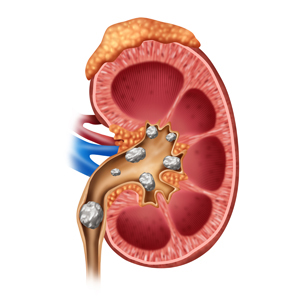
Each kidney has three layers – the outer layer (cortex), inner part (medulla), and central pelvis. Blood flows into the cortex for filtering. The medulla absorbs substances to return to the bloodstream. Waste is taken by tubes to the central pelvis, a collecting area where urine is emptied out into two tubes called ureters, and then passes to the bladder.

Multi-purpose organs

The two kidneys sit high in the back the abdomen. Each one is about the size of a fist, shaped like a bean, and surrounded by a protective layer of tissue.

Adrenal gland

The adrenal glands are small glands located on top of each kidney. They produce hormones that you can’t live without, including sex hormones and cortisol. Cortisol helps you respond to stress and has many other important functions. Adrenaline released from this gland makes the heart beat faster in scary situations.

Renal artery

The renal arteries are responsible for carrying oxygen-rich blood to your kidneys, which in turn help the kidneys rid your body of waste and excess fluid. This artery carries blood into the kidney to be filtered.

Renal vein

The renal vein is an asymmetrically paired vessel that carries the deoxygenated blood from the kidney to the inferior vena cava. Cleaned blood is carried from the kidney by the renal vein.

Outer casing

The kidneys and adrenal glands are wrapped in a layer of fat and strong outer tissue. The outermost layer is a tough connective tissue layer called the renal fascia.

Central pelvis

Urine collects here, and is then sent to the bladder. The pelvis, which is shaped somewhat like a funnel that is curved to one side, is almost completely enclosed in the deep indentation on the concave side of the kidney, the sinus.

Renal cortex

The cortex is the outer part of the kidney. It contains the glomerulus and convoluted tubules.

The renal cortex is surrounded on its outer edges by the renal capsule, a layer of fatty tissue. Together, the renal cortex and capsule house and protect the inner structures of the kidney.

Renal medulla

This layer of the kidney absorbs water, making urine more concentrated. The renal medulla is split up into a number of sections, known as the renal pyramids. Blood enters into the kidney via the renal artery, which then splits up to form the interlobar arteries.

Left ureter

This is one of two tubes that carry urine down to the bladder. There are two ureters, one attached to each kidney. The upper half of the ureter is located in the abdomen and the lower half is located in the pelvic area. 
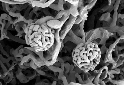
Nephrons

The kidneys contain tiny blood-filtering units called nephrons. Each nephron contains a glomerulus, a bundle of blood vessels surrounded by a capsule. As blood passes through the glomerulus, waste and excess water ooze into the capsule and are carried away by a tubule (tiny tube). Any useful substances, such as glucose, are absorbed by capillaries, while the waste is carried away to form urine.


Picture Credit : Google

What is the function of kidney?

Your two kidneys filter and clean the blood by removing toxic chemicals like the heart, the kidneys are at work every second of every day, producing a continuous flow of clean blood.

As blood circulates it picks up waste substances produced by the body’s cells. These would poison you if they were not removed from the body. The kidneys extract the toxins and excess water from the blood and process them to make urine. As well as cleaning the blood, the kidneys also release hormones, stimulate red blood cell production and keep the body’s water content balanced.

They release more urine if you have drunk a lot and less if you are dehydrated.


Picture Credit : Google