The female skeleton is usually smaller and lighter than a male’s with a wider pelvis, which makes childbirth easier.
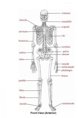
Cranium
This contains and protects the brain, eyes, ears, and nose. The interrelationships formed by neurons to create working memory, the plasticity demonstrated when adapting to injury, and the process of sorting through the wealth of visual and auditory stimuli contained in our world are all amazing feats unto themselves. The brain, which performs these various functions, is protected by a part of the skull called the cranium. We’ll turn our attention to the eight bones that form it: the ethmoid bone, the sphenoid bone, the frontal bone, the occipital bone, two parietal bones, and two temporal bones.
Lower jaw bone (mandible)
The only part of the skull that can move is the mandible. In anatomy, the mandible, lower jaw or jawbone is the largest, strongest and lowest bone in the human facial skeleton. It forms the lower jaw and holds the lower teeth in place. The mandible sits beneath the maxilla. It is the only movable bone of the skull (discounting the ossicles of the middle ear).
Spinal column
A flexible series of bones holds the head and upper body upright. The Spinal Column is also called the vertebral column. The bones in the spine are called vertebrae (ver-ta-bray). The column starts at the base of the skull and continues to the pelvis. Alternate layers of bone (vertebrae) and cartilage (car-til-ledge, the intervertebral discs) stack vertically one on top of the other in the spinal column. The lattice-like structure of the cancellous bone (cancel-lus, the spongy interior) in a vertebra absorbs external pressure.
Clavicle
This long bone is also called the collarbone. The clavicle (collarbone) extends between the manubrium of the sternum and the acromion of the scapula. It is classed as a long bone, and can be palpated along its length. In thin individuals, it is visible under the skin.
Scapula
Also called the shoulder blade, connects the arm to the shoulder. In humans they are triangular and lie on the upper back between the levels of the second and eighth ribs. A scapula’s posterior surface is crossed obliquely by a prominent ridge, the spine, which divides the bone into two concave areas, the supraspinous and infraspinous fossae.
Sternum
Also called the breastbone, it supports the ribs at the front of the body. The sternum (or breastbone) is a flat bone located at the anterior aspect of the thorax. It lies in the midline of the chest and has a ‘T’ shape.
As part of the bony thoracic wall, the sternum helps protect the internal thoracic viscera – such as the heart, lungs and oesophagus.
Humerus
This is the upper arm bone. It is located between the elbow joint and the shoulder. At the elbow, it connects primarily to the ulna, as the forearm’s radial bone connects to the wrist.
Ribs
The 12 pairs of curved rib bones protect the heart and lungs. The ribs partially enclose and protect the chest cavity, where many vital organs (including the heart and the lungs) are located. The rib cage is collectively made up of long, curved individual bones with joint-connections to the spinal vertebrae.
Ulna
This is the inner bone of the forearm. The ulna is located on the opposite side of the forearm from the thumb. It joins with the humerus on its larger end to make the elbow joint, and joins with the carpal bones of the hand at its smaller end.
Pelvis
These connected bones support the abdominal organs. The pelvis consists of four bones: the right and left hip bones, the sacrum, and the coccyx. The pelvis has several important functions. Its primary role is to support the weight of the upper body when sitting and to transfer this weight to the lower limbs when standing.
Radius
This is the outer bone of the forearm. It lies laterally and parallel to ulna, the second of the forearm bones. The radius pivots around the ulna to produce movement at the proximal and distal radio-ulnar joints.
Carpal
There are eight of these small bones at the wrist. Carpal bone, any of several small angular bones that in humans make up the wrist (carpus), and in horses, cows, and other quadrupeds the “knee” of the foreleg. They correspond to the tarsal bones of the rear or lower limb.
Metacarpal
There are five of these bones in each hand, at the base of the fingers and thumb. The metacarpals together are referred to as the ‘metacarpus.’ The tops of the metacarpals form the knuckles where they join to the wrist.
Phalanges
There are 14 phalanges in each hand, forming the fingers and thumb. The phalanges are the bones that make up the fingers of the hand and the toes of the foot. There are 56 phalanges in the human body, with fourteen on each hand and foot. Three phalanges are present on each finger and toe, with the exception of the thumb and large toe, which possess only two.
Femur
The largest leg bone is also called the thighbone. It functions in supporting the weight of the body and allowing motion of the leg. The femur articulates proximally with the acetabulum of the pelvis forming the hip joint, and distally with the tibia and patella to form the knee joint.
Patella
This bone is the kneecap. It is a small, freestanding, bone that rests between the femur (thighbone) and tibia (shinbone). The femur has a dedicated groove along which the kneecap slides. As a form of protection, both bones also contain cartilage — strong, flexible tissue — in the areas near the patella.
Tibia
The front of this bone is the shin. It forms the knee joint with the femur and the ankle joint with the fibula and tarsus. Many powerful muscles that move the foot and lower leg are anchored to the tibia.
Fibula
The smaller bone of the lower leg, this is located alongside the tibia. It runs parallel to the tibia, or shin bone, and plays a significant role in stabilizing the ankle and supporting the muscles of the lower leg. Compared to the tibia, the fibula is about the same length, but is considerably thinner.
Tarsal
There are seven of these small bones at the ankle joint. The tarsus is a cluster of seven articulating bones in each foot situated between the lower end of the tibia and the fibula of the lower leg and the metatarsus. It is made up of the midfoot (cuboid, medial, intermediate, and lateral cuneiform, and navicular) and hindfoot (talus and calcaneus).
Metatarsal
These five long bones sit between the tarsals and phalanges. Metatarsals are part of the bones of the mid-foot and are tubular in shape. They are named by numbers and start from the medial side outward. The medial side is the same side as the big toe.
Picture Credit : Google